Sesamoiditis
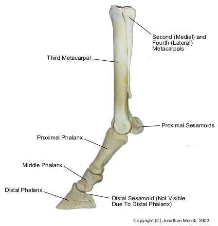
Sesamoiditis describes inflammation within the sesamoid bones at the back of the fetlock joint. The sesamoid bones have a similar action to the navicular bone. They act as 'pulleys' for the suspensory ligament as it passes over the back of the fetlock joint.
Sesamoiditis is most common in racehorses, eveners’ and show jumpers where high loading and over flexion of the fetlock joint results in tearing of the sesamoid ligaments and internal bone stress leading to sesamoiditis. Both front legs are usually affected.
Overweight young racing and sport horses, particularly fillies and horses with long pasterns and low heels, appear to be more prone to development of inflamed sesamoids. It may have an inherited risk, similar to navicular disease.
The proximal sesamoid bones in the horse are two small bones sitting at the base of the cannon bone in back of the fetlock joint. They are critical components of the suspensory apparatus that supports the horse's distal limb. The suspensory ligament originates from the bottom of the carpus (knee) or tarsus (hock) and runs down the back of the cannon bone, attaching to the top of the proximal sesamoid bones. The distal sesamoidean ligaments originate from the bottom of the proximal sesamoid bones and run down the back of the pastern, attaching at the back of the long and short pastern bones. The suspensory ligament, proximal sesamoid bones, and distal sesamoidean ligaments make up the suspensory apparatus of the fetlock joint and work together to prevent overextension of this joint when the limb is fully weight-bearing. Because the proximal sesamoid bones are integral in the suspensory apparatus of the distal limb, they cannot be successfully removed.
Sesamoiditis refers to pain associated with the proximal sesamoid bones. It often results from inflammation at the interface of the suspensory ligament and distal sesamoidean ligaments with the sesamoid bones. It is caused by unusual strain to the fetlock joint. It is most often seen in racehorses and jumpers, but it can be seen in any breed or discipline of horse.
Signs of sesamoiditis include lameness that often worsens when the horse is worked on hard surfaces. It can be exacerbated by fetlock joint flexion tests. There may be heat, swelling, or firm thickening around the fetlock joint, and the horse might be sensitive to palpation of the sesamoid bones.
A definitive diagnosis of sesamoiditis is made by taking radiographs (X rays) of the fetlock joint and sesamoid bones. Radiographic changes include increased size and number of vascular channels within the bone, calcification (hardening) of soft tissue structures surrounding the bone, proliferative bony production on the sesamoid bones, and avulsion (separating) fractures at the attachments of the suspensory ligament and distal sesamoidean ligaments to the sesamoid bones. If sesamoiditis is diagnosed based on radiographs, it is important to also ultrasound the suspensory ligament and
distal sesamoidean ligaments to assess any damage they might have.
Treatment goals include reducing inflammation in the sesamoid bones and fetlock joint. Rest is crucial until the horse is sound at the trot, at which point he can be built back up slowly. In the initial stages of inflammation, alternating hot and cold therapies, and poultices on the fetlock, will help reduce inflammation. Non-steroidal anti-inflammatory drugs such as phenylbutazone work systemically to decrease inflammation. Intra-articular treatment of the fetlock joint with hyaluronic acid will locally reduce inflammation in the joint.
Correct shoeing is critical to managing sesamoiditis. Some horses do well in egg bar shoes with squared toes and a wide roll or bevel. Having the toe pulled back and setting the shoe back will ease breakover, thus reducing stress on the suspensory apparatus. Other therapies such as blistering and extracorporeal shock wave therapy have been used with varying degrees of success.
Horses often need up to eight months of convalescence before returning to full work. Prognosis varies greatly depending on severity and whether surrounding ligaments are affected.


